Analysis Aids in Heart Valve Design
Simulation of prosthetic heart valves has become so essential that researchers have invented a Latin phrase for it: in silico.

June 1, 2010
By David Essex
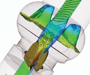 This CFD velocity profile of the middle plane of an ANSYS FLUENT model shows the downstream blood-flow velocity (with iso-lines in black) of a prosthetic aortic heart valve. Abaqus was also used to model the valve itself. The superficial tetrahedral grid is shown with the valve’s two leaflets in gold. |
While the artificial heart holds sway over the imagination as the penultimate component to perfect for saving lives, the less romantic—but no less amazing—reality consists of small prosthetic devices intended to replace failing sections of the heart and surrounding vascular system. With scaffold-like, artery-clearing stents now commonplace, the new vanguard is replacement heart valves.
Open-heart surgery, often risky, can now often be avoided with minimally invasive “percutaneous” procedures that deliver valves via catheters inserted through an artery, typically in the groin. The latter technique is standard for stents.
In a 2006 study, Millennium Research Group predicted the U.S. heart valve market would grow to $700 million, thanks to exploding development of mitral and aortic percutaneous valves and increased incidence of heart disease among the elderly.
Medical devices usually go through in vivo testing on live animals or in humans participating in clinical trials, and in vitro in the controlled environment of a test tube or petri dish. But increased use of desktop design tools has given birth to a new Latin phrase: in silico.
That’s because computer design, simulation, and modeling tools enable speedier introduction of safer, more durable valves.
Planned Interventions
Academic researchers and leading valve makers, including Edwards Lifesciences and Medtronic, have focused much of their computer work on designing the devices and simulating their operation. But imaging technology is also playing a prominent role in the ancillary efforts of marketing and training.
For example, the University of Colorado Denver used soft-tissue imaging such as computer tomography (CT) and magnetic resonance imaging (MRI) to develop 3D models of individual patients’ hearts and cardiovascular systems. The files are then converted to physical models on a Z Corporation 3D printer at the nearby Protogenic division of Spectrum Plastics Group. Doctors then use the model to plan and practice the procedure. “We can mimic very realistic patient cases, so we can train surgeons on complicated cases,” says James Chen, a UC Denver associate professor of medicine who helped develop the technology. One manufacturer, ValveXchange, has licensed the technology, and Chen says others are using patient data sets created by the university to test their devices.
Nearby Medical Simulation Corp., whose medical director is Dr. John Carroll, a co-inventor of the UC technology, makes simulated models that it claims are used by 95 percent of valve makers.
“What we do for companies like Edwards is design simulations around their products,” says Sammy Peppers, the company’s director of business development. “Operators actually experience the forces—the haptics—of what they will be doing.” Peppers says the company uses Adobe Photoshop and Autodesk Maya along with custom software for its simulations, while making the physical models from valve prototypes. It doesn’t need the level of detail that designers do, but takes information from the valve development process, as well as CT scans and other medical images from actual patients, to write programs that simulate live procedures. The method purportedly trims time to market and training expenses.
“With the physical model, you can really practice the intervention,” Carroll says. “A lot of this is done in fluoroscopy—they’re basically looking at live X-rays. What ]surgeons are] looking at is what they would actually see in the cath lab.”
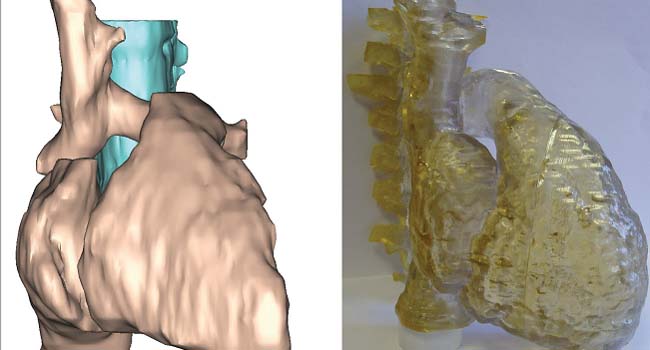 Rapid prototyping is commonly used to create physical models of the heart to aid in valve implantation. Researchers used medical imagery to create the 3D model of a pediatric patient’s heart (pink) and spine (blue) at left, which they then used to build the polymer model at right. |
Hagop Kaneboughazian, animation director at Viscira, uses Luxology’s modo software to make 3D models that manufacturers use to market their devices to doctors and investors. “We show how the model works inside the heart,” Kaneboughazian says. “It also has to fit within the anatomy. It has to be believable.” Still, the animation can’t be so blood-and-guts realistic that it might turn off non-physicians, he says.
To get the animations right, Kaneboughazian typically meets with the manufacturer’s chief science officer. “Half the time we start from sketches or conceptual drawings,” he says, though sometimes CAD files are available.
Kaneboughazian says modo is the initial modeling tool, while the animation is done in either NewTek’s LightWave or Autodesk Softimage. Then he brings the project back into modo for texturing, lighting, and rendering.
CFD: The Vital Flow of Life
Benchtop tests use physical models to simulate a valve’s behavior in fluid. Developers and academics supplement them with computer simulations. Chen says UC mostly uses ANSYS FLUENT and Simulia’s Abaqus to perform computational fluid dynamics (CFD) and finite element analysis (FEA). They use Autodesk Maya for smoothing of boundary information and assurance that boundaries don’t intrude on each other. Files are converted to stereolithography (STL) format for export to the Z Corp printer. McNeel Rhino software is also used for 3D modeling, according to Chen, while modo smoothes surfaces where sparse data points create over- or under-segmentation, a serious error that can confuse chambers of the heart. The university also employs several CAD programs.
Simulating Heart Valves on CILEA’s High-Performance Computers To simulate the hemodynamic (blood flow) characteristics of St. Jude Medical’s Hemodynamic Plus valve, the Italian high-performance computing consortium, CILEA HPC, used an impressive array of raw CPU power and off-the-shelf software. According to engineer Raffaele Ponzini, CILEA’s computer cluster was ranked 135th-fastest in the world in 2008. It is composed of 208 two-way Intel Xeon 3.16GHz QuadCores with 16GB of RAM per node and a total disk capacity of 13TB. The operating system is Red Hat Enterprise Linux Server Release 5.1, and James River Technical’s PBS Professional is used to manage workloads. Ponzini says the team used an ultra-fast cinematographic technique to acquire patient data to build a CFD model with realistic fluid dynamics that took into account heartbeats per minute, cardiac output, and the geometry of the valve housing and hinges. CILEA also studied the valve’s effect on blood platelets. “The CFD model, already equipped with the FSI algorithm, has been enriched with a particle-tracking algorithm to provide fluid-dynamics data of passive tracers,” which Ponzini says played the role of platelets. “We ran several simulations performing 4,000 time steps per heart cycle,” Ponzini says. “The CFD model was fully developed to be deployable on a parallel machine architecture, and thanks to the scalability properties of the problem, we achieved our results on a single heart cycle in about two days, performing the computations on up to 16 cores.” Ponzini says modeling and mesh generation were done in ANSYS GAMBIT software, and simulations were performed in ANSYS FLUENT by adopting a control-volume-based technique to solve the Navier–Stokes equations. — DE |
Keith Perrin, Autodesk’s industry principal, describes a “hive” of valve development centering on his company’s platforms, principally Maya, which he says is especially suited for modeling organic material. Inventor is preferred for valve design, and Algor Simulation for analysis, or to translate CFD into FEA. Perrin says some companies also use Autodesk software to create Method of Application (MOA) animations for training, typically by exporting Inventor designs back into Maya.
One of the main areas of study involves using CFD to predict how valves will interact with, and sometimes injure, the body’s soft tissue. Perrin says turbulent flow is important to model because it can show where blood flow might be affeccted by the valve. Developers also study laminar flow, which some designs deliberately harness to open and close a valve’s leaflets or flaps. “You can actually model where that boundary layer is going to break down,” Perrin says. “In order to understand the flow going across the valve, you need the complexity of the body immediately around it. You can’t just simulate the valve in isolation.”
Researchers are also studying how impact with prosthetic valves can damage the blood’s platelets or cause clots, according to Thierry Marchal, industry director for healthcare and consumer products at ANSYS. “The flow rate can be too small, and it can cause the platelets to stick,” Marchal says. “It can lead to death.” So researchers have done much work in FLUENT to study the phenomenon.
“A detailed description of the local hemodynamics is necessary,” explains Raffaele Ponzini, an engineer at CILEA HPC, an Italian research consortium for high-performance computing. For three years, Ponzini, who also does CFD analysis for yacht design, has worked on a team performing vendor-independent in vitro and in silico tests of St. Jude Medical’s Hemodynamic Plus valve (See sidebar).
FEA of Stress on Hearts
The flow of blood and pressure from soft tissue can cause a device to fracture, with obviously dire consequences. FEA is often the tool that pinpoints stress points before a bad design reaches patients.
Subham Sett, life sciences industry lead at Simulia, says valves “are under constant cyclic load. That leads to a lot of fatigue issues because of the opening and closing of the valves.” Maximum and alternating stresses are the most critical points in predicting when a valve might break, he says.
University of Connecticut and University of Pittsburgh researchers use Abaqus extensively to study soft-tissue behavior, especially valve leaflets to see if they close completely, according to Sett. “What we have within Abaqus is tools which let people solve flow problems in which there is a lot of structural influence.” He notes that Abaqus doesn’t do full-fledged CFD, but has multi-physics integrations to other third-party tools that do, such as ANSYS FLUENT and CD-adapco’s STAR-CD.
Like stents, the percutaneous soft-tissue valves are rolled up into tight cylinders that can fit inside the vein or artery, then expanded at the implant site. Shape memory alloys, which can “remember” the device’s previous shape, are increasingly popular materials. Many simulation studies have focused on the stress placed on valves when they’re rolled up.
The Next Frontier: Live Procedures
There is emerging interest in using some of the same computer imaging that was used in development to guide doctors while delivering the valves by catheter.
Chen says the UC hospital cath lab is already outfitted with the necessary imaging equipment, but it’s up to surgeons to use it. The main challenge is integrating the different imaging formats that are needed to present the complete picture in real time. Fluoroscopy is the only way to see the catheter, but it is 2D and can’t adequately show soft tissue. One promising approach is to combine it with images from 3D echocardiography.
Marchal agrees that unifying the images is a major research challenge. “The CT scan can export some geometry into STL format,” he says, “but it t usually needs quite a lot of repair.” ANSYS has begun a three-year venture with Medtronic’s Invatec division and an Italian university to speed up simulations so doctors can see the effects of interventions as they happen. “When the patient is lying on the table, you can’t wait for a simulation,” he says.
More Info:
ANSYS
Freelance writer David Essex has covered information technology for 24 years. He was a BYTE editor and has written for Computerworld, PC World, and numerous other publications. Send e-mail about this article to [email protected].
Subscribe to our FREE magazine, FREE email newsletters or both!
About the Author
DE’s editors contribute news and new product announcements to Digital Engineering.
Press releases may be sent to them via [email protected].