Dentistry in 3D Is Something to Smile About
A new virtual world of design and manufacturing brings dental decision-making to dentists and patients online.

August 27, 2008
By Susan Smith
 3D printing and digital imaging bring a new virtual world to the dentistry profession. Dr. Stephen M. Schmitt of Voxelogix Corp., San Antonio, TX, said in a presentation given at RAPID 2008 in May, that the future of dentistry would embody tools to allow surgeons to join digital datasets together, and to allow patients to go online and make design decisions regarding their teeth.
There are lots of reasons to keep teeth, and there are even more people who may lose their teeth as the current U.S. “baby boomer” population ages, whom Schmitt numbers at 30 million. Regardless of which way patients go, “Most people have no predictable way to fix their teeth,” noted Schmitt. They suffer from periodontal disease and dry mouth as a result of the medications they take. Dental surgeries can be painful and do not always reap positive results.
Dental surgeons consider that, although expensive, implants provide a better alternative to the removal of teeth and dentures, because the resultant bone loss as a result of teeth removal will be significant.
The issues that Schmitt sees in the dental industry are that dental technology is highly intensive labor. Much of it is done at the artisan level, by hand. Secondly, implant treatment is poorly integrated and prohibitively expensive for most people. Dr. Schmitt said he would like to see the cost of implants drop to under $40,000, the price of a moderately priced car as opposed to $75,000, as is the current outlay.
A potential solution to these challenges would involve the following:
- Unified digital data set
- Online treatment planning
- 3D visualization of planned treatment
- Direct digital fabrication of titanium parts
 Today most surgeons do not have any knowledge of STL files, or know how their systems work, said Schmitt, but can put a 2D file in a patient’s file and use it for a consultation.
With a server-based viewer process, the data would be loaded onto the server, and a physician could interact with it online through a viewer (to avoid the firewall problem). He or she could then make this available to a patient so they can view the choices. This could subsequently reduce the costs of appointments.
Diagnostic procedures
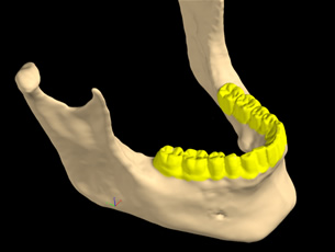
Digital imaging using a dataset from a CAT Scan and data model can be used for making upper and lower impressions, said Schmitt. “The CT Byte allows us to join two datasets together, to make a precise model to manufacture either using RP, milling or traditional dental processes.” To make an impression, a dental model is scanned and goes from ASCII text to DXF to an STL file.
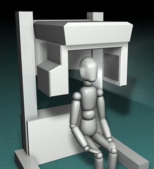
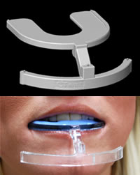
Medical CT scan or Cone Beam CT scan is now a common procedure that is lower cost and can be done in a dental clinic rather than a hospital. These procedures require reformatting in DICOM format and can take numerous images. One process employs taking facial photos for an accurate representation of the patient’s smile, to ensure that after surgery, the patient retains their smile. These photos show where the muscles attach when the patient smiles, important information that can’t be procured from a CAT Scan.
For people who have no teeth, a CAT Scan does not produce a clean image, it produces scatter, which makes it hard to put teeth in the right place. “With CT Byte, we can remount the casts, reproduce the biting position of the patient and move the virtual model by moving the physical model and joining the two models together,” Schmitt explained.
 In DICOM, the skull, the teeth of the upper and lower jaw, can be manipulated. A Boolean operation is done to separate the teeth from bone, and an implant can be completed in one day. “We extract the teeth, put implants in where they’re going to go, and compress treatment to one major surgery and one minor one a few months later,” said Schmitt.
With the virtual tools, surgeons can put new teeth in with existing teeth and in relationship to lips as well as to bone. If there is a need to reshape bone, the surgeons need to know that before surgery. The process involves only minimal pain.
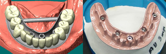
At a three-month period, the surgeon takes imaging of the soft tissue bridge as it has healed, and then builds a substructure made of titanium underneath that the implants attach to. The teeth are completed first. The titanium is covered with pink material so that when the artificial teeth are attached, the mouth looks natural. The STL file is translated in two file formats — CNC and STL, an example of “hybrid manufacturing.”
Electron beam melting can be used to create one-off custom parts and geometries that can’t be built any other way.
Benefits/value
Summarizing the benefits of using digital imaging and STL, Schmitt recounted the following: reduced costs, integrated treatment plan, pre-treatment visualization of outcome, and healthcare providers maximize skills.
Schmitt also said that we can now grow bone, and that we have come a long way in bone regeneration.
Overall, the combination of digital imaging, CAD, the Internet, digital manufacturing and new materials give dentists the opportunity to dramatically improve the options available for their patients.
Voxelogix Corp.
San Antonio,TX
Susan Smith is a contributing editor for Desktop Engineering magazine. Send e-mail about this article to [email protected].
Subscribe to our FREE magazine, FREE email newsletters or both!
About the Author
DE’s editors contribute news and new product announcements to Digital Engineering.
Press releases may be sent to them via [email protected].