Using Patient-Specific Data as a Basis for Design, 2 in a Series
Integrating anatomical STL files with rapid technologies to create 3D reconstructions is still difficult to accomplish.


Latest News
May 3, 2009
By Susan Smith
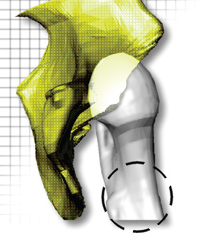 Reconstruction of CT image data of a young patient with congenital hip dysplasia (approx. 50 slices). Data courtesy of Shriners Hospital, Minneapolis, MN and Image3, LLC. |
The intersection of CAD and rapid manufacturing with medical anatomical scans remains tricky to navigate. At the forefront of finding a smoother integration is Alair Emory, who heads up digital design and development for Javelin 3D, a maker of anatomical 3D reconstructions and medical models based in Park City, Utah.
Emory, who holds both chemical engineering and biology degrees, bought her first rapid-prototyping machine in 1991. She was also a founder of Javelin 3D, Image3, and BodyReplicas, as well as a co-developer of Velocity 2 software, a package that imports grayscale image data to create 3D surfaces.
In a presentation at RAPID 2008, titled, “Integrating Anatomical STL Files with CAD,” Emory said her goal was to use patient-specific data as a basis for design. “I have clients who would like to be able to use this data in their CAD program,” she said. “They’re only asking for use of part of the spine, to be able to cut and drill holes in the spine and fit their devices to these geometries. This is still very difficult, expensive to do, and in some cases, not possible.”
Emory said her company uses software that will import medical scan data like CAT scans for bone. Javelin 3D’s Velocity2 software enables identification of the anatomical region of interest and then “links each slice together in 3D to reconstruct the identified region.” CAT scan data is not considered 3D data for CAD packages.
By using Velocity2, designers can achieve an STL with Dicom, Tiff, or other proprietary 2D grayscale image data, and do segmentation, 3D reconstruction, smoothing, and file-size reduction all within the same program. Emory said there are other programs that can do the same thing, such as Able Software’s 3D Doctor and Mimics from Materialise.
Once 2D grayscale data is imported, designers can seek the tissue they’re interested in reconstructing within the data. The problem, said Emory, is that medical reconstruction is not well integrated with rapid manufacturing technologies.
“Often you’re working with a radiologist or surgeon who only knows about a Dicom format, nothing else,” she said. A multidisciplinary background is needed to be able to make the link between rapid technologies like 3D printing and medical product development. But there are other challenges as well: the Dicom format was standardized within the medical community, medical viewers are free, there are health insurance and litigation issues, and AMA standards and requirements must be met.
Emory suggests that the “Medical 3D Approach” to tackle these challenges incorporates obtaining the smallest possible file size without losing important details; finding ways to communicate geometry details across multiple disciplines (e.g., using STL file viewers); and defining CAD conversion software requirements that are necessary to drive medical applications forward.
“I used a sample STL file I had reconstructed from CAT scan data—18.2 megabytes,” said Emory. In evaluating the products, she approached vendors directly or downloaded software in demo, shareware, or freeware formats.
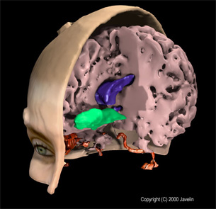 Composite of structures in human brain as reconstructed from MRI data3D Files were exported from Velocity2 PLUS and image was rendered via third- party software. Image courtesy of Javelin. |
STL File Viewers
Emory said she looked for STL file viewers that were easy to install, able to handle large files, included measurement features, were low in cost or free, could print 2D views, and ran on “basic” PCs. She explained that printing 2D views was important because the medical community can easily view them. Among her favorites from the research, Emory said MiniMagics by Materialise topped the list. Other choices include shareware such as Quick3D, which has “confusing zoom control but good customer service,” and a product called i-Know 2D from CCIM, which easily generates 2D technical drawings and measurements but is not freeware.
Anatomical File Size Reduction
For medical rapid prototyping, file-size reduction should be done directly in the package used to generate the file, according to Emory. “If you’re working in medical RP you shouldn’t use secondary file-size reduction programs.”
There are some legal reasons for this. “When you go to third-party software packages that do STL file reduction, you don’t get input back in general as you do in the medical RP package,” explained Emory. Also, she said, you shouldn’t get rid of the medical data form file. The industrial engineer may want to do that, otherwise, how would the engineer determine what the most important aspect of the anatomy was?
Because anatomical files are detailed, if you get a medical STL file, you need to tell the person doing the reduction what your needs are. Emory said it was important to reduce the file sizes very carefully.
“If you are working with an industrial part … it would be common for the service bureau to run the industrial file through a file size reduction operation before building the prototype,” she said. “The service bureau wants to reduce the file size because large files can be difficult to pre-process for the RP build operation. With an industrial part it would be pretty easy for the RP operator to visually look at the original file and a size-reduced file, and give the go-ahead to process the build.
“In general,” she said, “medical STL files are large and the concern is that the RP service-bureau operator is automatically going to want to reduce the file size. However, what might not seem important to the RP operator might actually be important to the medical diagnostician.… It seems that the best option would be to create a standard for medical STL files that restricts file modifications to the originator of the file.”
An example of a stand-alone reduction is VizUp, which can also serve as a viewer.
CAD Programs
CAD programs used commonly for anatomical STL include SolidWorks and PTC’s Pro/ENGINEER. Rhino from McNeel North America is also very capable. The recommendation from SolidWorks technical support is that anatomical STL files be processed by third-party software conversion programs before importing into SolidWorks.
Rhino was “most responsive,” according to Emory. “Technical support claims import into their program without problems, and we were able to demonstrate import of vertebra files into the Rhino software.” In spite of this, Emory said they did not have any clients using Rhino.
Pro/ENGINEER has a reverse-engineering module and contains licensed code for surface fitting, which is a kernel within the base code. “We were able to put together a demonstration for numerous modifications to a jaw STL file provided for a dental webinar,” said Emory.
Conversion Programs
According to Emory, conversion programs are costly but provide valuable solutions. SolidWorks recommended Geomagic software (to work with anatomical STL file conversion to NURBs you should only need two modules and a specific graphics card); NuGraf from Okino with many output options; and Rapidform from INUS Technology.
Emory finished by saying that the area of medical RP is still very confusing to a lot of people. Some programs simply don’t work and solutions are still expensive. She suggested that standards be developed for the use of medical RP data. Her suggestions include:
- Guidelines on medical RP file modifications
- Guidelines on conversion of these files to other file formats for use with standard CAD programs or FEA analysis
- Guidelines on quality of original medical scan data
- Recommendations on appropriate use of RP systems (Some RP systems are much more accurate than others but medical practitioners can get confused by all the options.)
- Recommendations on use of 3D physical medical models in the operating room. RP models have been used regularly outside the U.S. in the operating room.
More Info:
Able Software Corp.
Lexington, MA
CCIM
Leiden, The Netherlands
Geomagic
Research Triangle Park, NC
INUS Technology, Inc.
Seoul, South Korea
Javelin 3D
Park City, UT
Materialise
Leuven, Belgium
McNeel North America
Seattle, WA
Okino Computer Graphics
Toronto, ONT.
PTC
Needham, MA
SolidWorks
Concord, MA
VizUp, Inc.
Vancouver, B.C.
Susan Smith is a contributing editor for Desktop Engineering magazine. Send e-mail about this article to [email protected].
Subscribe to our FREE magazine, FREE email newsletters or both!
Latest News
About the Author
DE’s editors contribute news and new product announcements to Digital Engineering.
Press releases may be sent to them via [email protected].